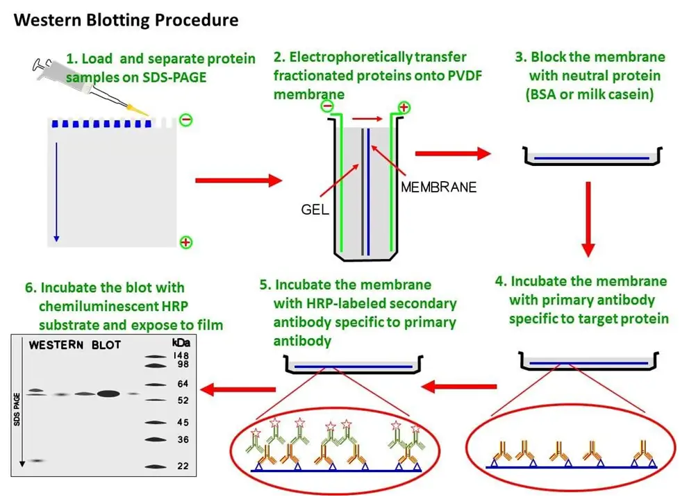
The increased sensitivity of conjugated secondary antibodies, compared to primary antibody only, results from these antibodies binding to the primary antibody at multiple locations, which amplifies the signal. Remember to check that the light emission wavelength of a conjugate is compatible with your reading device/scanner. Using an enzyme-linked secondary antibody, such as horseradish peroxidase (HRP) or alkaline phosphatase (AP), or a western blot-optimized fluorescence conjugate (eg IRDye ®), offers a high level of sensitivity. To visualize your protein, you need to select a secondary antibody that will bind to the primary antibody.

In this video, you’ll learn a few useful tips for choosing a primary antibody for western blot. If possible, choose a primary antibody that has been knockout (KO) validated to ensure it binds specifically to the intended target. You should select a primary antibody raised in species different from that of your sample. The sensitivity and specificity of your western blot depend on the quality of the antibodies used. In this video, we’ll recap the essential steps of western blot and show you a specific example of the interpretation of western blot results. The membrane can be further processed with antibodies specific for the target of interest and visualized using secondary antibodies and detection reagents. Protein transfer to the membrane is essential because gels used for electrophoresis provide a poor surface for immunostaining, ie antibodies don’t stick to the proteins in the gel. The gel is then placed next to the membrane, and the use of an electrical current induces the proteins to migrate from the gel to the membrane. In the first step, the proteins are separated based on size by gel electrophoresis.

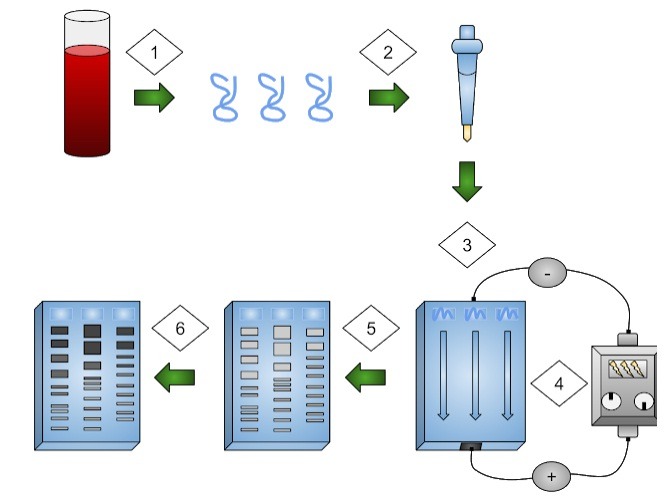
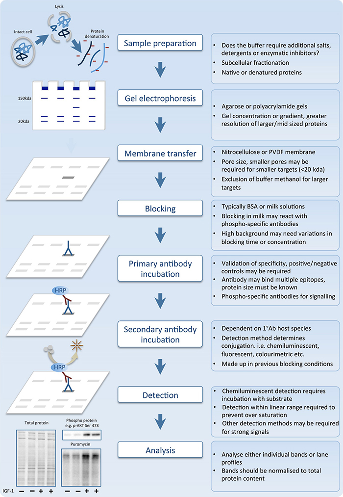
To achieve this, western blot implements three steps: (1) separation by size, (2) transfer to a solid support, and (3) visualizing target protein using primary and secondary antibodies.

Western blot allows us to determine the relative protein levels between samples and establish the molecular weight of the target, which can provide insight into its post-translational processing. Western blot, or western blotting, is a technique widely used in research to separate and identify proteins. You’ll get to grips with the essential information required before launching into the protocol itself.
#Western blot steps series
In Part 1 of our series on western blot, we’ll take you through the basics of this incredibly popular and useful technique. We’ll guide you through western blot basics and essential protocols before moving on to optimization, troubleshooting, and more advanced techniques. Search our extensive portfolio of primary antibodies and secondary antibodies specifically designed for western blotting.Welcome to our training series on western blot. The secondary antibody is then visualized by staining, immunofluorescence, radioactivity, or other methods. A secondary antibody (an antibody to the primary antibody) is then added (to bind to the primary antibody). The blot is covered with a buffer that contains an antibody that will bind to specific parts of the protein being studied. Following separation by electrophoresis, and then transferred (blotted) from the gel to a membrane for specific visualization. A protein sample is denatured to prepare it for gel electrophoresis.

The western blot (WB) is a common protein detection and analysis method. Steps include: sample preparation, gel electrophoresis, membrane transfer, antibody probe, detection, imaging, and analysis. Kits, buffers, blocking agents, reagents, membranes and substrates for use in conducting western blotting and ELISA immunoassays includes accessories, modules, and replacement parts and solutions.


 0 kommentar(er)
